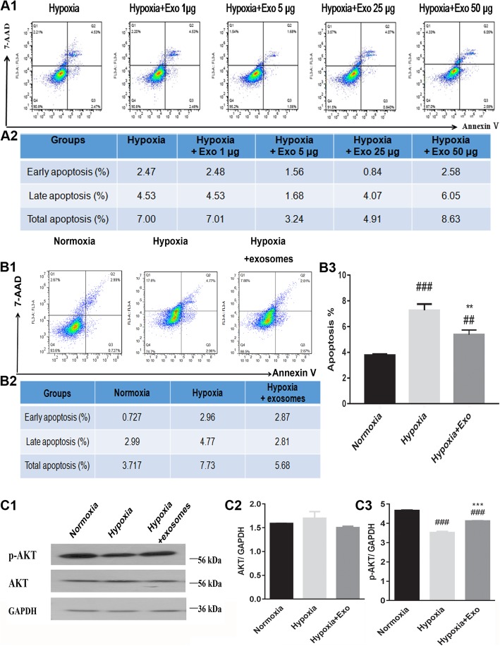
Fig. 3.
Mesenchymal stem cell-derived exosomes affected H9C2 cell apoptosis and p-AKT/AKT expression levels when cultured in hypoxic conditions. A1, A2 Increasing concentrations of exosomes had an effect on the amount of H9C2 cell apoptosis in hypoxic growth conditions. B1–B3 Flow cytometry analysis showed that exosomes protected H9C2 cells from apoptosis in hypoxic growth conditions. C1–C3 Exosomes regulated the expression levels of p-AKT and AKT in hypoxic growth conditions. ###P < 0.001 vs. normoxia; ##P < 0.01 vs. normoxia; ***P < 0.001 vs. hypoxia; **P < 0.01 vs. hypoxia. Exo, exosomes; p-AKT/AKT, phosphorylated protein kinase B. Statistics calculated based on the results of three repetitions of each experiment

