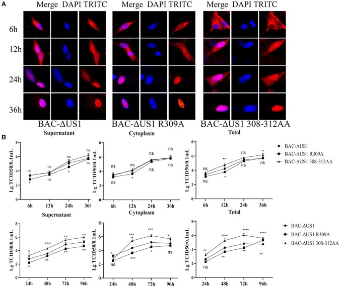
Figure 10.
Intracellular localization of ICP22 and viral titres of one-step and multi-step replication kinetics. (A) Intracellular localization of ICP22 in infected DEFs at different time points with 10 MOI BAC-ΔUS1, BAC-ΔUS1 R309A, and BAC-ΔUS1 308-312AA. (B) Viral titer in the cytoplasm, supernatant, and total of one-step growth assays (top panel) and multi-step replication kinetics (bottom panel). Each time point was measured in triplicate, with the standard error indicated. A representative experiment with at least three repeats was performed. Ns represents no significance, *p < 0.1, **p < 0.01, ***p < 0.001 and ****p < 0.0001.

