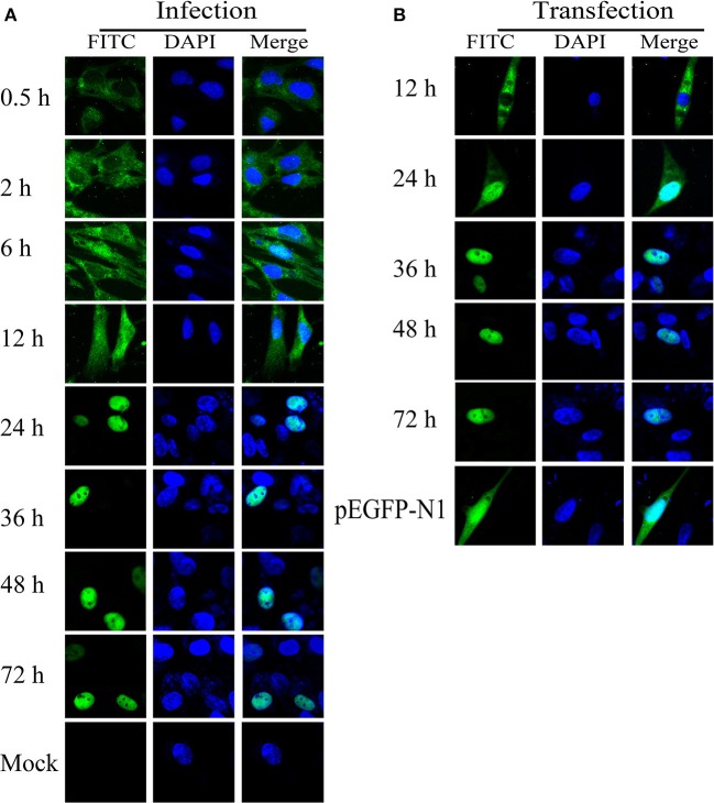
Figure 8.
Intracellular localization of ICP22 in DEFs. (A) Intracellular localization of ICP22 in infected DEFs at different time points with 10 MOI; mock was used as a control. (B) Intracellular localization of ICP22 at different time points in EGFP-ICP22-transfected DEFs. pEGFP-N1 was used as a control.

