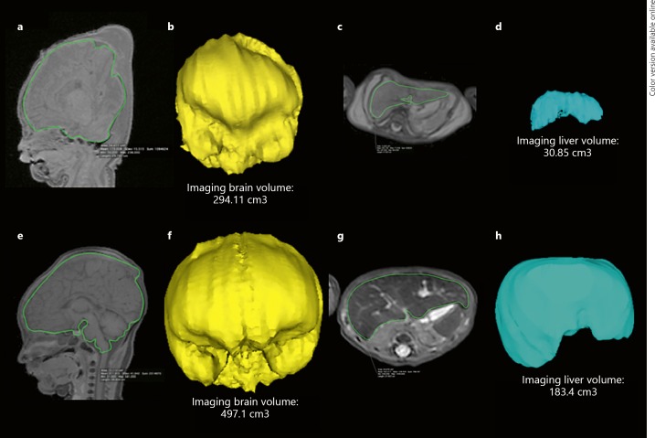
Fig. 1.
PMMR of a stillborn 39-week gestational age fetus with FGR (a–d) and a stillborn 40-week non-FGR (control) patient (e–h). MRI manual segmentation on isovolumetric T1-weighted sequences of the brain are shown in sagittal views (a, e, green outline) with corresponding derived 3D volumes (b, f). A similar method was adopted on isovolumetric T2-weighted imaging of the abdomen for the liver in axial sections (c, g, green outline) with corresponding derived 3D volumes (d, h). For the fetus with FGR (a–d), the IB: LvR was calculated as 9.53 (AB: LwR was 7.6), and for the control fetus without FGR (e–h) the IB: LvR was 2.7 (AB: LwR was 2.4).

