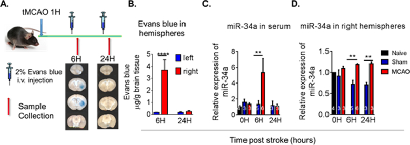
Figure 2. Time course of miR-34a following ischemia in mouse serum and brain following stroke.

(A) Focal cerebral ischemia was induced by tMCAO for 60 minutes in C57BL/6 male mice and Evan’s blue was injected into the mice (i.v.). Transcardiac perfusion was then performed and brain images acquired as shown by representative coronal brain sections. (B) Quantification of Evan’s blue extravasation in the left and right hemispheres. Data are expressed as mean ± S.D.; n=4/group; One-way ANOVA followed by post-hoc Tukey’s test, ****p<0.0001. (C) At the indicated time post-stroke, tissue samples were collected and miRNA levels in the ischemic brain hemisphere and serum were analyzed by qRT-PCR. In mouse serum following tMCAO, miR-34a levels were increased by 6 hrs post-stroke. (D) miR-34a levels were increased in the brain of tMCAO mice by 6 hrs and at 24 hrs. Data are expressed as mean ± S.D., **p < 0.01. (Naive: N = 2, Sham: N = 3, tMCAO: N = 3) [Jun et al., 44]
