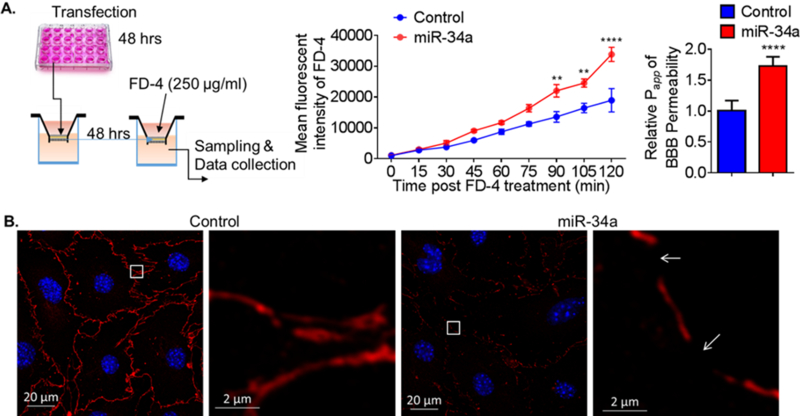
Figure 3. Overexpression of miR-34a increases BBB permeability in vitro.

(A) A schematic protocol using fluorescein isothiocyanate–dextran-4 (FD-4) to detect BBB permeability in vitro. FD-4 permeability in CECs that overexpressed miR-34a plasmid (0.017 ng) versus control was presented as real-time rate of FD-4 mean fluorescent intensity (2-way ANOVA followed by post hoc Dunnett’s test; N = 3; **, p < 0.01; ****, p < 0.0001). Calculated apparent permeability coefficient Papp (Student’s t-test; ****, p < 0.0001) is expressed as mean ± SD. (B) Confocal fluorescence images of confluent monolayers confirmed microscopically after transfection with miR-34a plasmid versus control. Fluorescent staining: tight junctions ZO-1 (red), cell nuclei (DAPI, blue). Overexpression of miR-34a apparently disrupted tight junctions and resulted in gaps between cells (white arrows). Results are representative of three independent experiments. [21]
