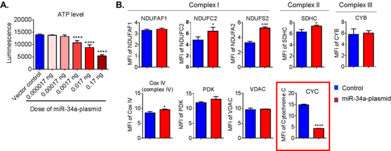
Figure 4. Overexpression of mir-34a reduces mitochondrial function and decreases CYC level in cerebrovascular endothelial cells.

(A) ATP level was measured at 72 h post-transfection. Data are expressed as mean ± SD (N = 5). One-way ANOVA followed by post hoc Tukey’s test. (****, p < 0.0001). (B) Flow cytometry analysis of mitochondrial specific proteins for complex I proteins (NDUFAF1, NDUFC2 and NDUFS2), complex II protein (SDHC), complex III protein (CYB), complex IV protein (CYC C oxidase, Cox IV), CYC, pyruvate dehydrogenase kinase (PDK), and voltage-dependent anion channel protein (VDAC) at 72 h post-transfection. CYC level was significantly lower in the cells that were transfected with the miR- 34a plasmid. Data are presented as mean ± SD (N = 3) and analyzed by Student’s t-test, *, p < 0.05; ***, p < 0.001; ****, p < 0.0001. Results are representative of three independent experiments. [21]
