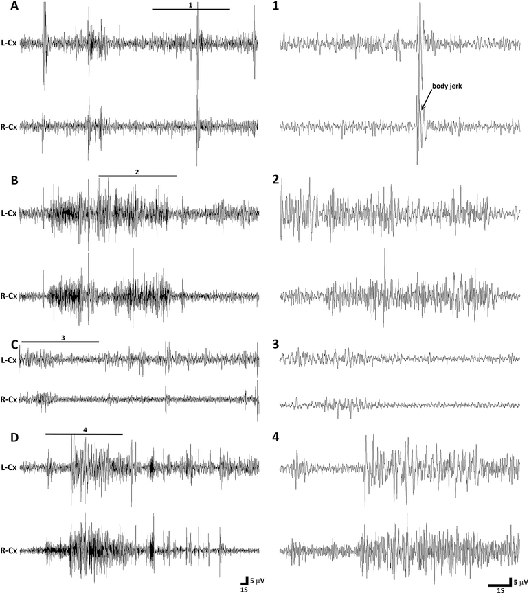
Fig. 2. Representative EEG tracings from P7 rats during immediate reperfusion period.
(A) The tracing on the left shows an example of inter-ictal EEG in a vehicle treated rat during the immediate reperfusion period. A magnified excerpt of a part of the EEG, marked with a bar above the tracing (1), is provided on the right side of the compressed EEG tracing. During this epoch the rat is not moving, except for some forelimb twitches and a whole body jerk preceded by a spike. (B) The tracing shows EEG activity during an electroclinical seizure that occurred approximately one hour in the reperfusion period in the same vehicle treated rat as above. The EEG ictal activity was associated with clonic movements of limbs. The enlarged excerpt of a part of the EEG (2) is provided next to compressed EEG tracing. (C) The representative tracing shows EEG activity in a flupirtine treated rat one hour in the reperfusion period. Except for the slight hindlimb movement at the start of the epoch, the rat was lying motionless during the entire epoch. The magnified excerpt of a part of the EEG (3) is provided on the right side of the compressed EEG tracing. (D) The tracing shows EEG activity during an electroclinical seizure in the phenobarbital treated HI rat. The EEG ictal activity was associated with tonic-clonic movement of limbs and thrashing-like motion of both hindlimbs. The enlarged excerpt of a part of the EEG (4) is provided next to compressed EEG tracing. L-Cx = left cortex, R-Cx = right cortex, S = second, V = volts.

