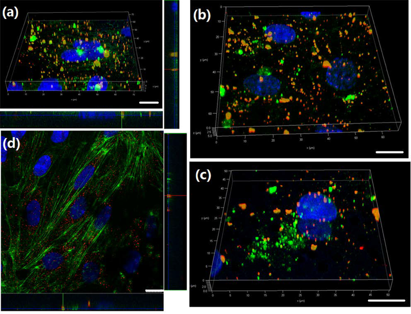
Figure 5.
(a) Representative 3D confocal image showed co-localization of PSi-Lf NPs (red) and EEs marker Rab-5 (green) on one-cell type BBB model. (b) Co-localization study of PSi-Lf NPs (red) with late endosome marker Rab-7a (green) on one-cell type BBB model. (c) Z-stacks reconstructed into 3D images of co-localization profiles of PSi-Lf NPs (red) with Lyso-Tracker (green) on one-cell type BBB model. (d) Co-localization study of PSi-Lf NPs (red) with Actin (green) on one-cell type BBB model. Scale bar: 10 μm.

