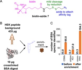
Figure 3.

Enrichment of crosslinked BSA from a complex sample. A) Structure of 7, with the biotin group (pink), the disulfide bond (green) and the azide moiety (blue). Upon reduction of the disulfide, the biotin moiety is removed from the crosslink. B) Depiction of the spike‐in experiment. 10 μg of crosslinked and CuAAC‐modified BSA digest were added to 435 μg of HEK digest as a complex background. Magnetic streptavidin beads were used to enrich the crosslinked peptides. C) Results of the HPLC‐MS2 analysis, showing the effect of enrichment. The experiment was performed in technical triplicates; numbers show the mean value and the standard deviation is indicated by the error bar.
