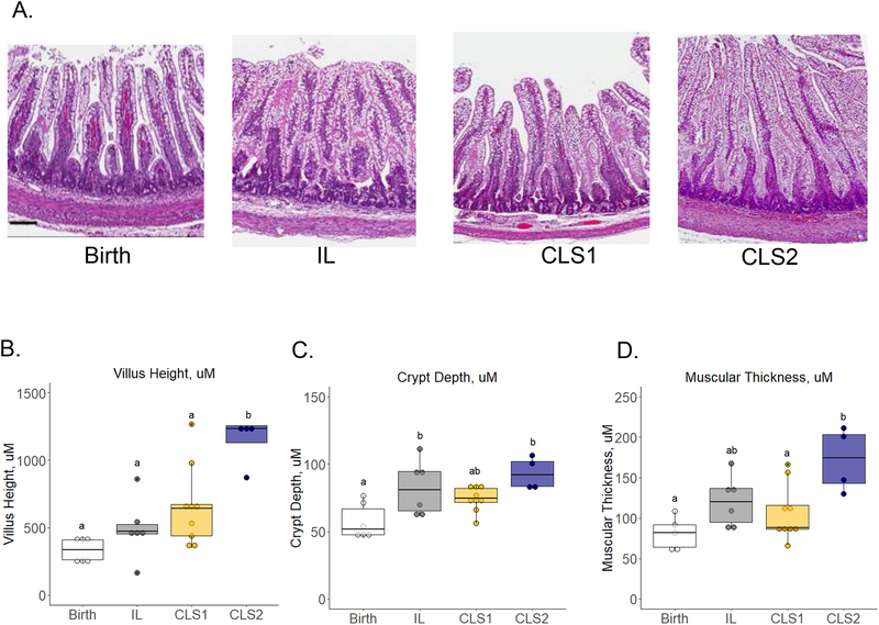
Figure 4.
Histologic evaluation of the distal ileum at birth (white) and in the IL (gray), CLS1 (yellow), and CLS2 (blue) groups. Representative hematoxylin and eosin-stained images at 5x magnification (A) are shown with quantification of villous height (B), crypt depth (C), and muscular thickness (D). Results shown as box plots. Labeled points without a common letter represent a statistically significant difference of P˂0.05. Scale bar =100um. CLS, concentrated lipid supplement; IL, Intralipid®-control.

