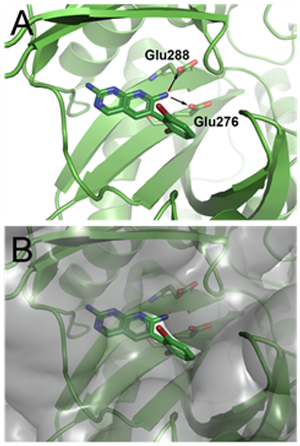
Figure 1.
Compound 1 (carbon, green; nitrogen, blue; bromine, dark red; all rendered as sticks) co-crystallized with E. coli BC at 2.1 Å as reported in Ref. 5 (PDB code 2V58). (A) View of compound 1 in the BC active site showing the 7-position vector that points toward two proximal glutamate residues as illustrated by the two arrows. (B) View of the solvent accessible surface representation, in transparent grey, of E. coli BC co-crystallized with 1. The second vector points off the dihalo-aromatic ring of 1 through a small slot in the active site surface toward solvent. Images prepared using PyMOL.

