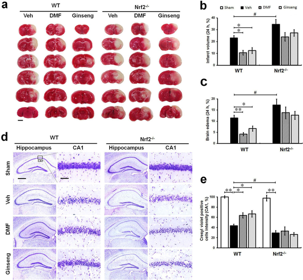
Fig. 1. Pretreatment with DMF or Ginseng reduces ischemia-induced infarct volume, brain edema, and hippocampal CA1 neuron loss 24 h after HI in WT mice, but not in Nrf2−/− mice.
(a) Representative images of TTC-stained brain sections (n=4–6 per group). (b and c) Quantitative analyses of infarct volume and brain edema. (d) Representative images of CV-stained hippocampus (n=3 per sham group and n=4–5 per ischemic group). (e) Quantitative analyses of hippocampal CA1 neurons indicated by CV staining. 24 h after HI, ischemia-induced infarct volume, brain edema, and hippocampal CA1 neuronal loss were significantly reduced in WT, but more importantly, not in Nrf2−/− mice pretreated with DMF or Ginseng, whereas Nrf2 deficiency significantly exacerbated the deterioration process. *P<0.05, **P<0.01, #P<0.05. TTC: 2,3,5-triphenyltetrazolium chloride; CV: Cresyl violet. Scale bar, 2 mm (a), 500 μm (d, hippocampus) and 50 μm (d, CA1 sub-region).

