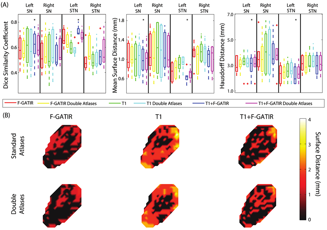
Figure 1:
Segmentation results for structures in the dienchephalon. Quantitative segmentation results are shown in (A). For the left SN, multi-modal segmentation with T1 and FGATIR outperformed other approaches (*; p<0.05; Wilcoxon sign-rank test). For the right SN no segmentation approach outperformed other approaches. For the left STN, multi-modal segmentation with T1 and FGATIR outperformed other approaches (*; p<0.05; Wilcoxon sign-rank test). For the right STN no segmentation approach outperformed other approaches. In (B), surface distances between the true and estimated segmentations for the left SN are shown for the six proposed segmentation approaches.

