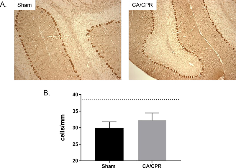
Figure 3.
CAMKIIα KO mice lack CA/CPR induced Purkinje cell loss. A. Representative images of calbindin labeling used to quantify Purkinje cell density in the cerebellum at 7 days after CA/CPR. B. Quantification of Purkinje cell densities at 7 days after surgery. CAMKIIα mice were subjected to sham surgery or CA/CPR. Dotted line represents Purkinje cell density in wildtype shams. N=4 per group.

