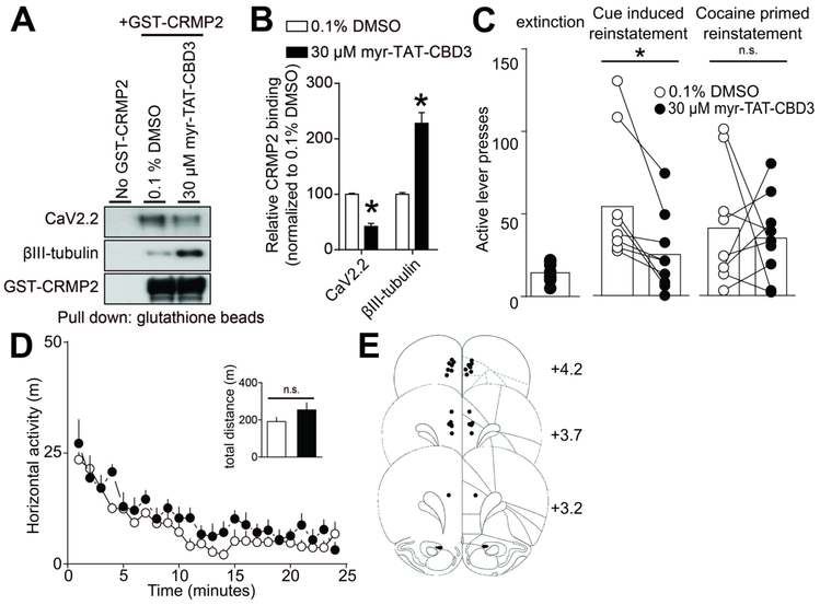
FIGURE 5:
Inhibition of CRMP2-CaV2.2 interactions in the mPFC decreases reinstatement of cocaine seeking in response to drug-predictive cues, but not cocaine-prime. (A) A glutathione (GST)-pull down assay and Western analysis illustrating the interaction of CRMP2 with CaV2.2 and βIII-tubulin. Brain lysates were incubated with either myr-TAT-CBD3 or control vehicle. Minimal non-specific protein binding occurs in the no GST-CRMP2 condition (lane 1). (B) Bar graph summarizing the actions of vehicle or myr-TAT-CBD3 on the binding of CRMP2 to CaV2.2 or control βIII-tubulin (*p<0.05, Mann-Whitney test.). The myr-TAT-CBD3 peptide effectively inhibited CRMP2 interaction with CaV2.2 in brain lysate (n=7). (C) Bar graphs summarizing lever press responses during (left) extinction or different types of reinstatement testing (middle, cued-reinstatement; right, cocaine prime [10 mg/kg, i.p.]). All reinstatement testing (2 hr) occurred following bilateral mPFC infusion of vehicle or myr-TAT-CBD3. Relative to respective infusions of vehicle, myr-TAT-CBD3 decreased lever pressing in response to cocaine-associated cues (t(8)=3.665, p=0.0064), but not cocaine-prime (t(8)=0.3768, p=0.7161). (D) Relative to a bilateral mPFC infusion of vehicle, the infusion of myr-TAT-CBD3 does not reduce the spontaneous locomotor activity associated with a novel environment. The summary time course illustrates horizontal photocell counts (in 1 min bins) collected during the initial 25 min period of a 2-hr locomotor test session started 10 mins following mPFC drug infusion. Inset bar graph represents the total (mean ± SEM) horizontal activity for the 2-hr session (t(8)=1.343, p=0.2162). (E) Atlas plates summarize the location of microinjection cannula tips (Paxinos and Watson, 1998). Numbers denote distance from bregma in the anterior-posterior plane. For all infusions, vehicle was 0.1% DMSO and myr-TAT-CBD3 was applied at 30 μM.

