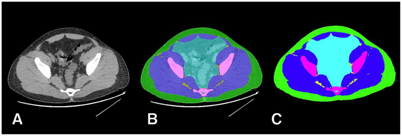
Figure 1.
Example of manual ground truth segmentation. The axial CT image at pelvic level (A) is manually segmented into multiple tissues (B) resulting in a mask (C) with 6 classes: background (black); subcutaneous adipose tissue (SAT, green); muscle (blue); inter-muscular adipose tissue (IMAT, yellow); bone (magenta); and miscellaneous intra-pelvic content (cyan).

