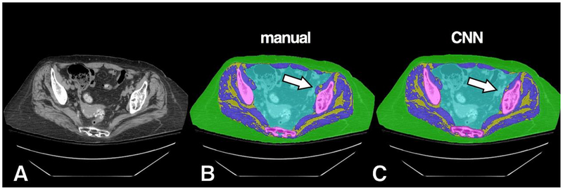
Figure 5.
Example of high accuracy segmentation with minor misclassification on a subject with more prominent IMAT. Original grayscale image (A), manual segmentation (B) and CNN segmentation (C). A small area of CNN misclassification is seen along the left iliopsoas muscle (arrow), which was predicted instead as intra-pelvic content.

