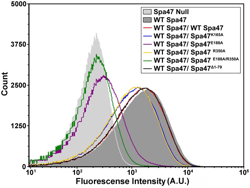
Fig 6. Flow cytometry fluorescence intensity histograms comparing extracellular T3SS injectisome tip protein presentation by Shigella strains expressing combinations of wild-type and mutant Spa47 constructs.
The tested Shigella strains were each treated with primary rabbit polyclonal antibodies against the T3SS tip protein IpaD and Alexa Fluor 647 conjugated goat anti-rabbit secondary antibodies to fluorescently label T3SS proteins on the bacterial surface. The histograms include 100,000 individual intensity measurements per condition and are representative of two independent biological replicates.

