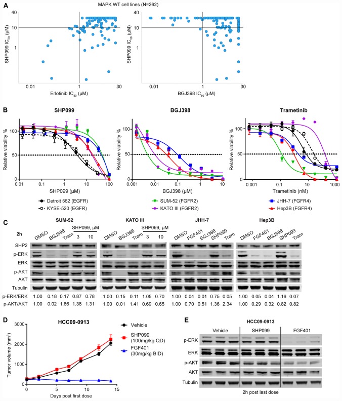
Figure 1. FGFR-driven MAPK-dependent cells are resistant to allosteric SHP2 inhibition.
(A) Correlation of anti-proliferation IC50 values of SHP099 and erlotinib or BGJ398 in 262 MAPK (KRAS, NRAS, HRAS, BRAF, and NF1) wild-type cell lines generated from high-throughput compound profiling as described [18]. Each dot represents one cell line. (B) Anti-proliferative effects of SHP099, BGJ398, or trametinib at the indicated concentrations in a 6-day cell proliferation assay with indicated cell lines (mean percentages of cell viability are shown, error bars denote standard deviation, n = 3). (C) Immunoblot of SHP2, p-ERK1/2 (T202/Y204), ERK1/2, p-AKT (S473), AKT, and tubulin with indicated cells treated with 0.5 μM BGJ398, 0.1 μM FGF401 for JHH-7 and Hep3B, 10 μM SHP099 (also 3 μM SHP099 for SUM-52 and KATO III), or 0.1 μM trametinib for 2 h. Ratios of p-ERK/ERK and p-AKT/AKT were calculated as described in Materials and Methods. (D) Mean tumor volumes of hepatocellular carcinoma patient derived xenograft HCC09-0913 in nude mice following treatment with vehicle, SHP099 (100 mg/kg body weight, daily), or FGF401 (30 mg/kg body weight, twice a day) for 14 days. Error bars, SEM. n = 3 mice per group. (E) Immunoblot of p-ERK1/2, ERK1/2, p-AKT, AKT, and tubulin from tumor tissues collected at 2 h after last dose from mice treated as described in (D).

