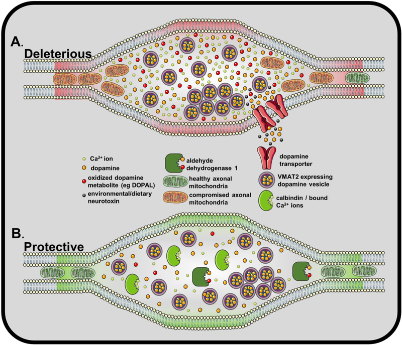
Figure 1. protective and deleterious elements of dopamine release sites.
A. Schematic of a release site in terminal regions of dopamine neurons. Release sites are flanked by unmyelinated axonal compartments, which harbor pools of axonal mitochondria. Repeated exposure to large increases in intracellular Ca2+ ions induce mitochondrial damage, leading to compromised bioenergetics. Cytosolic dopamine (orange) is hypothesized to be higher in disease sensitive neurons than non-sensitive counterparts, resulting in increased levels of oxidized dopamine metabolites (red) capable of causing damage to intracellular and transmembrane components. Additionally, dopamine transporters at release sites may uptake environmental and dietary neurotoxins at a level correlated with transporter expression. B. The Ca2+ binding protein calbindin, coupled with less reliance on Ca2+ for pacemaking, limits damage to axonal mitochondria preventing bioenergetic failure. Aldehyde dehydrogenase – an enzyme the converts aldehyde groups to carboxylic acid – acts on selected dopamine oxidation metabolites including dopamine -3,4,-dihydroxyphenylacetaldehyde (DOPAL) to reduce ROS generation and subsequent cell damage.

