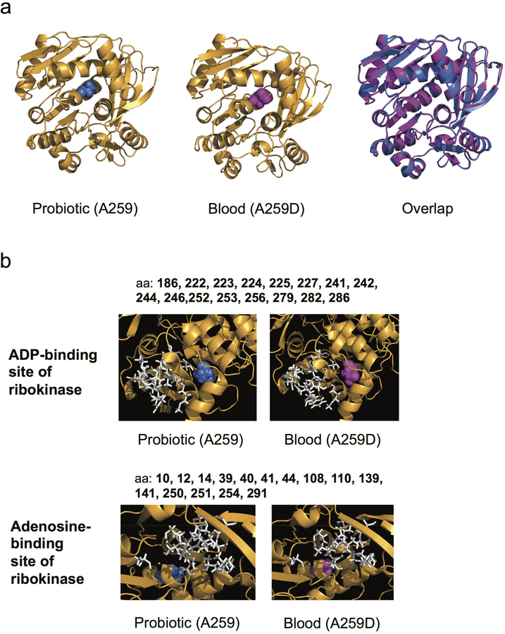
Extended Data Figure 3. The blood-isolate-specific ribokinase SNP does not perturb the predicted structure of ribokinase but occurs near the active site.
(a) Predicted structures of probiotic ribokinase with A259 (blue, left), blood isolate from Patient R1 with ribokinase A259D SNP (magenta, middle) and overlap (right). (b) The predicted binding site amino acids of ribokinase for adenosine are shown in white, with the alanine 259 (blue) of the probiotic (left) and the aspartic acid (magenta) of blood isolate 1 (right) shown compared to the adenosine-binding positions.

