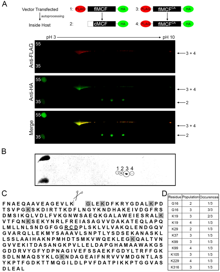
Figure 3.
MCF is N-terminally acetylated following auto-cleavage.
A. Anti-HA IP on cell lysates of HEK293T cells ectopically expressing dt-flMCF or dt-flMCFCA was individually completed. Samples were then mixed and run together for two-dimensional western analysis using IPG strips pH 3–10 with western blot completed using anti-FLAG (red) and anti-HA (green) antibodies. The mixing of samples helped to orient cleaved vs uncleaved fragments for the dt-flMCF sample. Red (top panel) and green (middle panel) channels were imaged separately and then merged (bottom panel). Numbers beside panels indicate which version of MCF in the schematic is found in each spot. B. Two-dimensional analysis with Coomassie gel was completed for only sample of dt-flMCF. Gel shows populations that were individually excised and analyzed by bottom up mass spectrometry. C. Protein sequence of MCF designating auto-cleavage site (dashed-line), and catalytic residues important for auto-proteolytic activity (underlined). Shaded residues are those found to be acetylated in populations 1–4. D. Table summarizing specific residues found to be modified and the number of times the modification was detected in each population.

