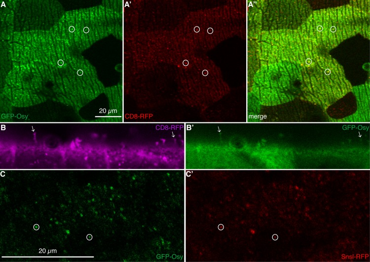
Fig 4. GFP-Osy localizes to pore canals.
By confocal microscopy, in live L3 larvae, GFP-Osy (green) is detected at the surface of epidermal cells marked by ridges in the top view (A). As described recently, these ridges are produced by the basal site of the cuticle engraved into the apical plasma membrane of epidermal cells of L3 larvae [38]. Occasionally, the GFP signal accumulates forming dots (circles). These dots co-localise (yellow) with red membrane-marking CD8-RFP dots (A’,A”). We observed that not all CD8-RFP dots coincided with a GFP-Osy signal; we suspect that there may be different CD8-RFP populations. Optical cross-sections confirm that CD8-RFP signals (magenta, B) protruding from the cell surface of live third instar larvae and marking pore canals coincide (arrows) with GFP-Osy signals (green, B’). In optical sections parallel to the surface, below the surface of the cuticle, dots of GFP-Osy signal are detected (C) that largely overlap with the signal of Snsl-RFP that marks the tips of potential pore canals (C’). Please note that images were taken sequentially of live animals, a perfect match of signals is therefore not expected.

