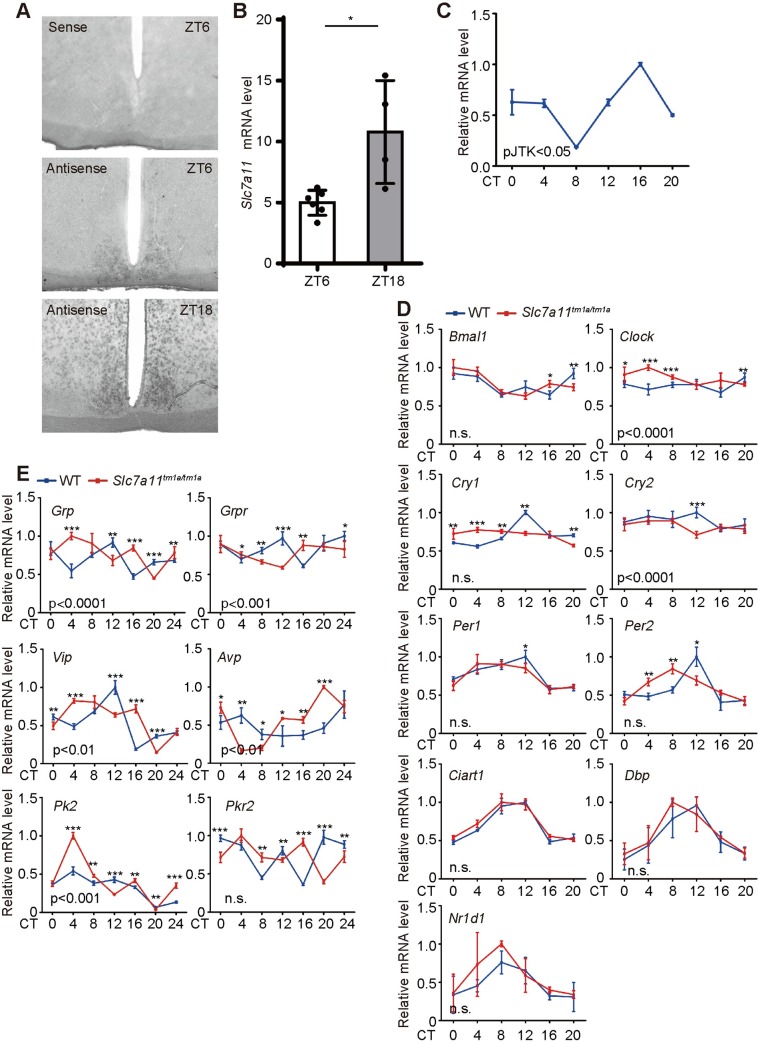
Fig 7. Altered expression of the coupling genes in the Slc7a11tm1a/tm1a mice.
(A) Expression of Slc7a11 in mouse SCN detected by in situ hybridization at ZT6 and ZT18. ZT, Zeitgeber Time. Coronal brain sections containing the SCN were hybridized with the cRNA sense (upper) or antisense probe (middle and lower) of Slc7a11 at ZT6 and ZT18. (B) Quantification of in situ hybridization signal of Slc7a11 by Image J from 3–4 coronal brain sections. *: p < 0.05. (C) Real-time PCR analysis of the expression of Slc7a11 in SCN of wild-type mice. Error bars represent the s.d. for each time point from three biological independent replicates. The rhythmicity of gene expression was determined based on the JTK algorithm (pJTK < 0.05). (D) Expression profiles of the core clock genes in the SCN from control and Slc7a11tm1a/tm1a mice. Also see the expression in the liver (S10 Fig). Error bars represent the s.d. for each time point from three independent replicates. (E) Expression profiles of the coupling factors in the SCN from control and Slc7a11tm1a/tm1a mice. Error bars represent the s.d. for each time point from three independent replicates. Two-way ANOVA was employed to test the statistical significance. *: P < 0.05; **: P < 0.01; ***: P <0.001.

