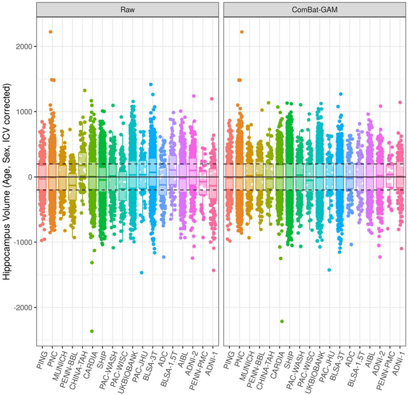
Figure 6:
Comparison of hippocampus volumes before and after harmonization, correcting for age, sex, and ICV using a GAM. Studies are ordered from youngest to oldest based on median age. In the left panel, volumes were not adjusted for site. In the right panel, volumes were adjusted with ComBat-GAM, which removes location (mean) and scale (variance) differences across sites after controlling for biological covariates. Horizontal lines are plotted at constants at 0, −200, and 200 for visual aid. Comparisons for additional ROI volumes are shown in Supplementary Figure 1.

