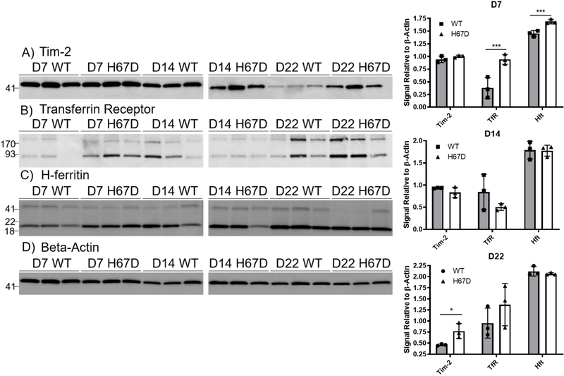
Figure 5. Tim-2, Transferrin Receptor, and H-ferritin expression during development.
Shown are representative western blots of expression for Tim-2 (A), TfR (B), FtH1 (C), and β-actin (D) on whole mouse brain homogenates. Western blots were repeated 3 times for each timepoint; each band represents homogenates pooled from 3 male and 3 female mice. E) Densitometric analyses of western blots reveal significantly elevated Tim-2 expression in H67D mice at day 22 and elevated TfR expression in H67D mice at day 7. For quantification we used the sum total of all of the bands because additional bands are thought to be dimers (near exact molecular weight matches). FtH1 expression was increased in H67D mice at day 7. Wild-type versus H67D means ± SD were evaluated for statistical significance by unpaired t-tests, n=3 biological replicates. *=p<0.05, ***=p<0.001.

