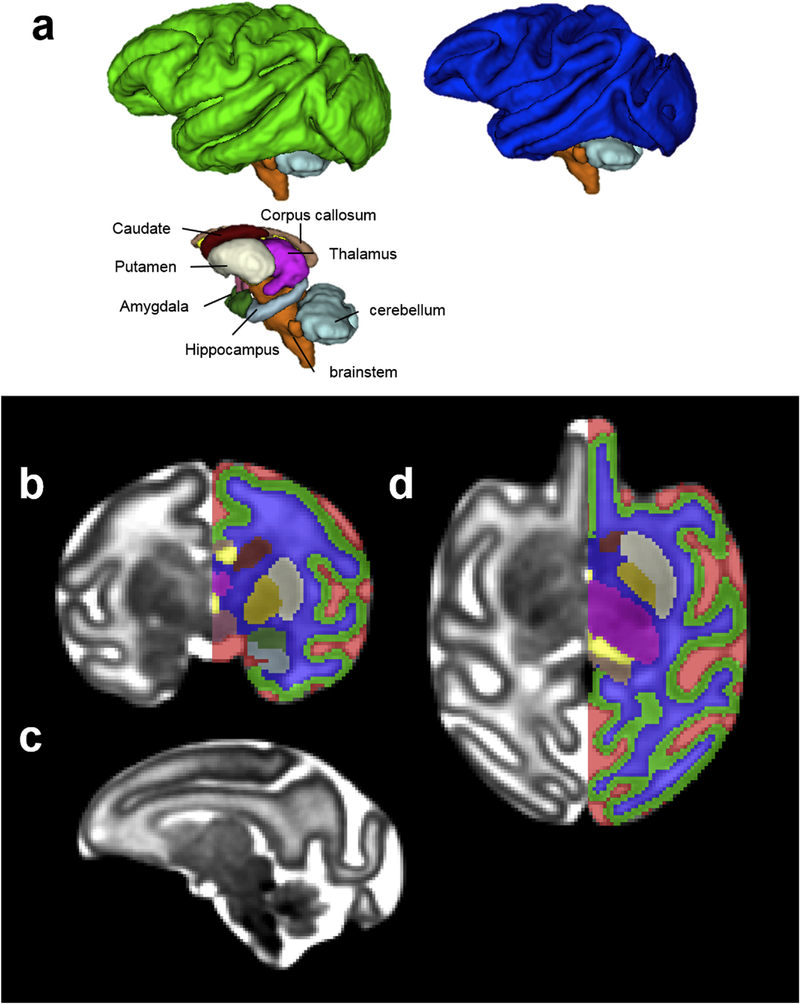
Fig. 3.
(a) 3D, (b) coronal, (c) sagittal, and (d) axial views of the G135 template and tissue segmentation. At G135, the lentiform nucleus can be further segmented into the putamen (cream) and the Globus pallidus (yellow) because of their increased contrast on T2-weighted images (b and d). At this developmental stage, the caudate nucleus appears hypointense compared to the putamen on the T2-weighted template (b and d). Although heterogeneity within thalamus can be observed (b-d), it is not further segmented into different nuclei. Likewise, brainstem is not further segmented into midbrain, pons, or medulla despite T2-weighted intensity differences (c).

