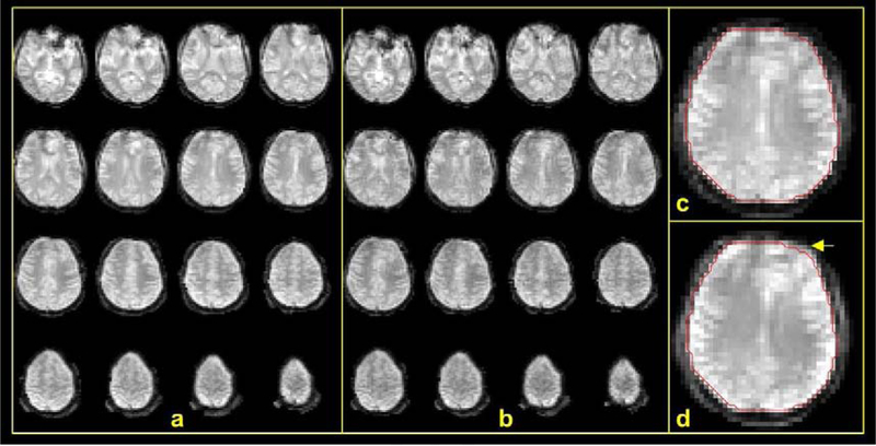
FIG. 2.
Quality of PN images. Shown are examples of PN images at 4 mm spatial resolution and temporal resolutions of 4.32 s and 0.54 s of the full (a) and fast (b) navigators, respectively. (a) Distortion-corrected full navigator images of the brain. (b) Distortion-corrected fast navigator images. (c) and (d) show the effect of distortion correction: (c) a slice of distortion-corrected image with red contour marking the brain boundary and (d) uncorrected image of the same slice. A yellow arrow points to the most notable difference of the brain boundary in the two cases.

