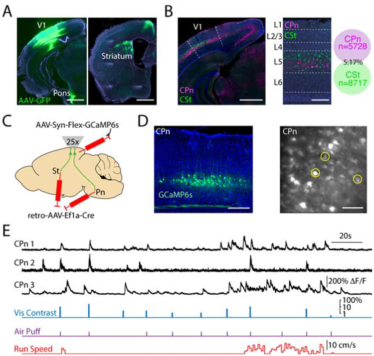
Figure 2. Two-photon imaging of activity in projection-specific layer 5 subpopulations.
(A) Injection of AAV5-hSyn-GFP into V1 (indicated by dashed lines) labels projections in both the ipsilateral pons (left) and ipsilateral dorsal striatum (right). Scale bar is 1 mm.
(B) Retrograde labeling of corticopontine (CPn, pink) and corticostriatal (CSt, green) cells with dual-color cholera toxin subunit B injections into ipsilateral pons or striatum reveals two non-overlapping populations in V1 (indicated by dashed lines) that are mostly limited to layer 5 (left, center). Scale bar is 200 μm. Co-localization of CPn and CSt neurons (right, n=3 mice).
(C) Schematic illustrating intersectional viral approach for conditional expression of the calcium indicator GCaMP6s in layer 5 PNs projecting to either the ipsilateral dorsal striatum (St) or pons (Pn).
(D) Left, example ex vivo image showing restricted expression of GCaMP6s in layer 5 corticopontine (CPn) neurons. Scale bar is 200 μm. Right, example in vivo 2-photon imaging of corticopontine neurons at 525 μm depth (image averaged over 5 seconds). Scale bar is 100 μm.
(E) Example traces showing simultaneous recordings of three CPn neurons from (D), timing of visual stimuli, air puff, and continuous running speed.

