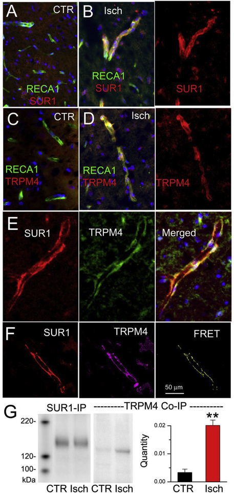
Figure 3. SUR1-TRPM4 is upregulated in microvessels post-ischemia.
A–D: Double immunolabeling of contralateral control (CTR) (A,C) and ipsilateral post-ischemic (Isch) (B,D) tissues for the endothelial marker, RECA1 (green), and SUR1 (red) (A,B), or RECA1 and TRPM4 (red) (C,D); superimposed images are shown in A; B, left panel; C; D, left panel. E: Double immunolabeling of ipsilateral post-ischemic tissues for SUR1 (red) and TRPM4 (green); superimposed images are shown at right. F: Double immunolabeling of ipsilateral post-ischemic tissues for SUR1 (red) and TRPM4 (magenta); FRET signal shown at right. G: Contralateral control (CTR) and ipsilateral post-ischemic (Isch) tissues underwent immunoisolation using anti-SUR1 antibody; the resultant isolate was immunoblotted using anti-SUR1 antibody (SUR1-IP) or using anti-TRPM4 antibody (TRPM4 Co-IP); bar graph: quantity of SUR1-TRPM4 Co-IP normalized to HSC70; 3 replicates; **, P <0.01.

