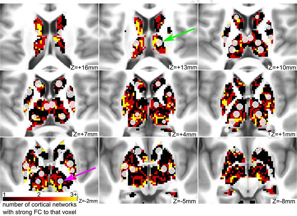
Figure 8: Some ROIs in the basal ganglia and thalamus have strong connectivity to multiple networks.
Integrative voxels in the basal ganglia and thalamus are displayed in red and yellow colors. These voxels have strong connectivity to more than one cortical functional network. Some of the novel ROIs (displayed in translucent white) contain a majority of integrative voxels (green arrow), while other ROIs contain few or none (purple arrow).

