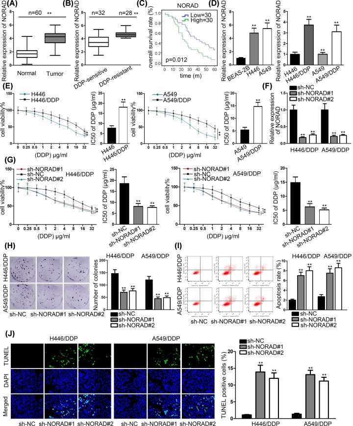
Figure 1. NORAD expression was lifted in NSCLC tissues and cells.
(A) RT-qPCR assays were used to test the expression of NORAD in NSCLC tissues and adjacent normal tissues. (B) NORAD expression was examined in DDP-sensitive and DDP-resistant tissues. (C) Kaplan–Meier curve was used to analyze the overall survival between high and low NORAD expression. (D) N0RAD expression was tested in NSCLC cell lines (H446 and A549) and normal human lung bronchial epithelial BEAS-2B cell as well as their DDP-resistant cell H446/DDP and A549/DDP cells. (E) MMT was conducted to evaluate cell viability of H446 and A549 while their parental cells were treated with DDP. (F) RT-qPCR assays were conducted to appraise the efficiency of sh-NORAD#1/2 in cells. (G and H) MTT and colony formation assays were conducted to examine the cell proliferation while H446/DDP and A549/DDP transfected with sh-NORAD#1/2 exposed to DDP. (I and J) Flow cytometry analysis and TUNEL were conducted to probe rate of apoptosis in H446/DDP and A549/DDP transfected with sh-NORAD#1/2 after exposure of 2 μg/ml DDP; **P < 0.01.

