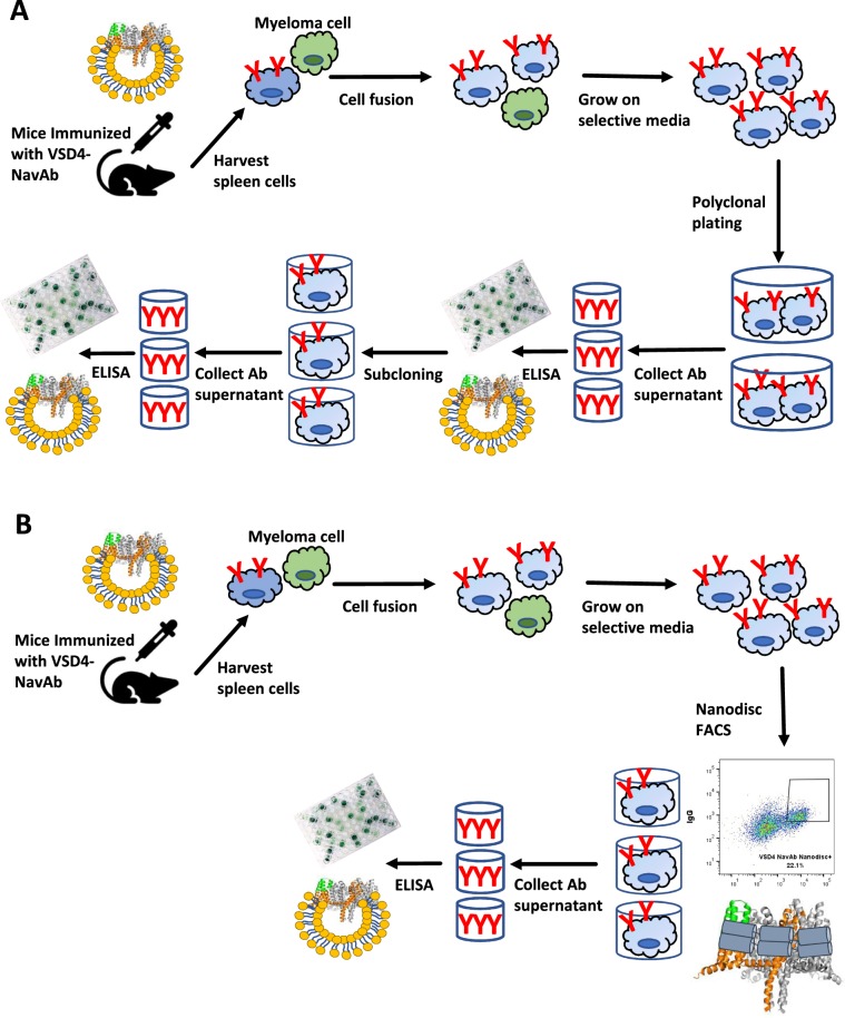
Figure 7.
FACS of hybridoma using antigen-incorporated nanodiscs facilitates antibody discovery. (A) Identification of monoclonal antibodies begins with the immunization of mice with antigen, followed by isolation of immune cells. Following fusion to myeloma cells, the hybridomas are grown and secrete antibody into the media for downstream screening. In the case of multi-pass membrane protein targets, where antigen-specific hybridomas can be very rare, and when soluble FACS reagents are not available for single cell sorting, the hybridoma are plated polyclonally and screening is performed on pools of antibodies. Upon successful identification of a well of interest, the hybridoma must then be subcloned to identify the individual hybridoma generating the antibody of interest. (B) Incorporation of the target of interest into nanodiscs (the two helical chains of MSP are shown as grey cylinders wrapping around the NavAb; lipid molecules are omitted for clarity) enables single cell FACS of hybridoma. This allows for the identification of antigen-specific hybridoma shortly after hybridoma fusion, thus reducing timelines and resources required to screen large numbers of polyclonal plates.

