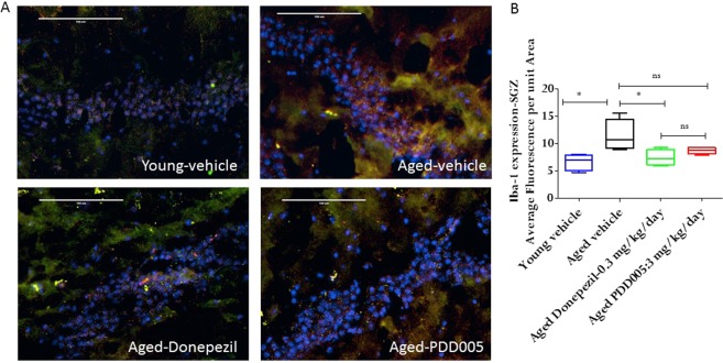
Figure 5.
IL-1 β synthesis, and microglial activation modulation in PDD005-treated aged mice. (A) Images showing IL-1β (green fluorescence), iba-1 (red fluorescence) expression. Yellow = Overlay; Blue = nuclei; SGZ region of mouse brain. 40X magnification, scale bar = 100 μm. (B) Graphs representing the average relative fluorescence of Iba-1 in SGZ. Significant differences determined by using one-way ANOVA with Tukey’s test. *P < 0.050, **P < 0.01 and ***P < 0.001 compared with aged-vehicle.

