Figure 8.
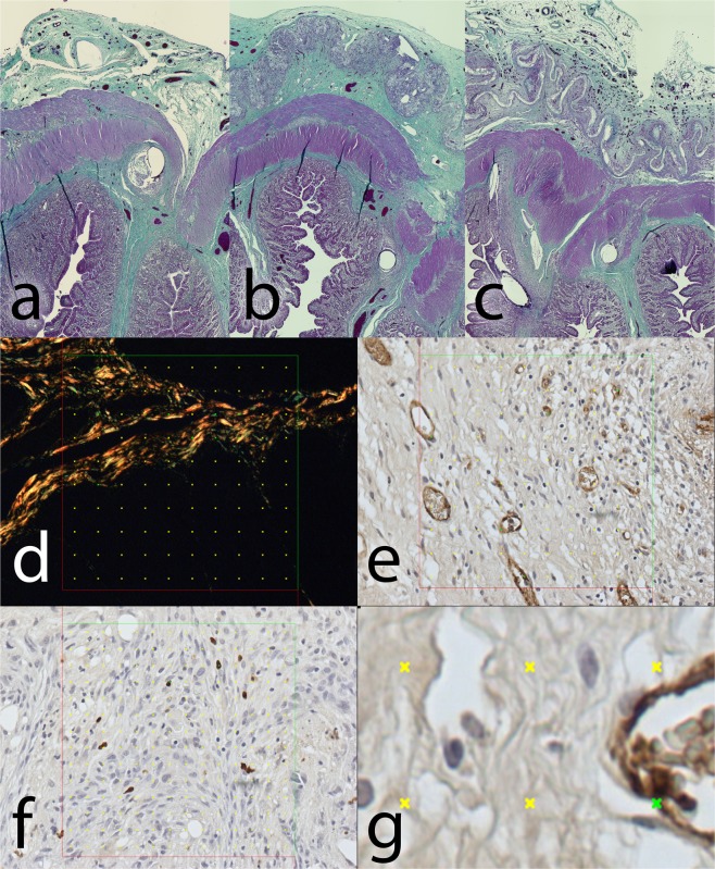
Histological staining of explanted anastomoses: (a) Green trichrome: Control group; (b) Green trichrome: PCL group, the empty space on the site of application of the nanomaterial can be seen in the upper layer, surrounded by normal granulation tissue; (c) Green trichrome: PLCL group, a much thinner empty area can be seen in the upper layer, also surrounded by normal granulation tissue; (d) PSR staining, collagen fibres stained yellow, stereological mesh; (e) vWF factor staining, the endothelial cells stained brown, stereological grid; (f) MAC 387 staining stereology, positive cells stained blue, stereological grid; (g) magnification of vWF staining stereology with a positive cross in the upper right corner.

