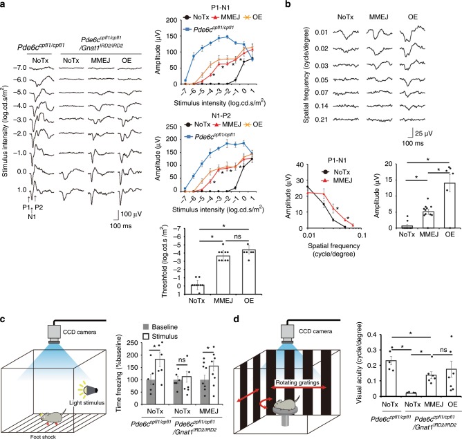
Fig. 3. Visual restoration by in vivo mutation-replacement genome editing.
Flash visually evoked potentials (fVEP) of the visual cortex contralateral to the eyes in response to flashes of various intensities. MMEJ indicates eyes treated with Gnat1 mutation replacement (N = 9) and OE (over-expression) indicates those with Gnat1 gene supplementation (N = 6), both delivered by single AAV. NoTx refers to untreated eyes (N = 9). Control Pde6ccpfl1/cpfl1 mice (N = 5). Note, light sensitivity as defined in the Methods was increased by ~4 log unit after MMEJ-mediated genome editing, which was not significantly different to the effect mediated by OE (right lower panel). b Pattern VEPs. N = 11, 10, and 6 for MMEJ, untreated and OE, respectively. c Fear conditioning test. Freezing time before (Baseline) and during (Stimulus) presentation of fear-conditioned light cue from MMEJ treated (N = 9) and untreated (N = 6) mice. d Optokinetic response. Note threshold of spatial resolution of vision (visual acuity) was not different in the MMEJ and OE. N = 10, 7, and 4 for MMEJ, OE, and NoTx, respectively. Control Pde6ccpfl1/cpfl1 mice (N = 6). Data represent the mean ± S.E.M.; *P < 0.05 (a, b, d, ANOVAs followed by Tukey’s post hoc test; c Student’s t-test); nd, non-detectable; ns, not significant. Source data are provided as a Source Data file.

