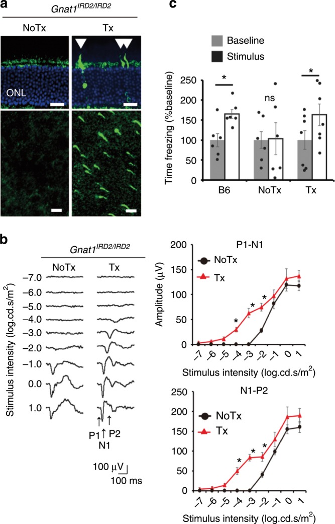
Fig. 4. In vivo mutation replacement genome editing in a mouse model of retinal degeneration.

a GNAT1-positive photoreceptors (arrowhead) following treatment of Gnat1IRD2/IRD2 mice shown in a retinal section (top) and a flatmount (bottom). Scale bar: 20 µm. b fVEPs recorded from contralateral visual cortices in treated and untreated eyes of the same mice. (N = 7). c Fear conditioning test, showing freezing time before (Baseline) and during (Stimulus) presentation of fear-conditioned light cue. Treated (Tx, N = 7) and untreated (NoTx, N = 6) Gnat1IRD2/IRD2 mice and CL57B6 mice (B6, N = 6). Data represent the mean ± S.E.M.; *P < 0.05 (Student’s t-test); ONL, outer nuclear layer. ns, not significant. Source data are provided as a Source Data file.
