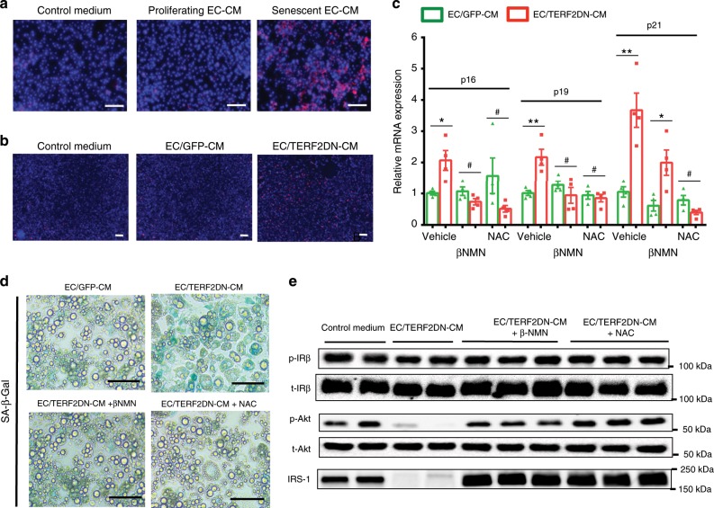
Fig. 3. Senescent EC impairs adipocyte functions by inducing oxidative stress.
a Superoxide was detected (as red fluorescence) in 3T3-L1 adipocytes treated with the control medium, or CM derived from proliferating young or replicative senescent EC. b Superoxide was detected in 3T3-L1 adipocytes treated with the control medium, or CM derived from control ECs (EC/GFP-CM) or premature senescent ECs (EC/TERF2DN-CM). c CDK inhibitor expression in 3T3-L1 adipocytes treated with the indicated EC-CM in the presence or absence of βNMN or NAC (n = 4 biologically independent samples each). A two-tailed Student’s t test was used for difference evaluation between the two groups. Data are presented as mean ± s.e. *P < 0.05, **P < 0.01, and #not significant. d SA-β-Gal staining in 3T3-L1 adipocytes treated with the indicated EC-CM in the presence or absence of βNMN or NAC. e Immunoblotting for the insulin signal pathway and IRS-1 in insulin-stimulated 3T3-L1 adipocytes treated with the indicated EC-CM in the presence or absence of βNMN or NAC. Bars: 100 μm. Source data are provided as a Source Data file.

