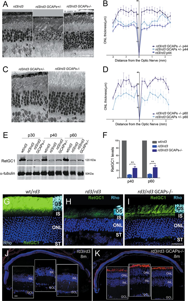
Fig. 2. Retinal degeneration in rd3 mice is substantially prevented by GCAPs ablation.
a Retinal morphology of rd3/rd3; rd3/rd3 GCAPs+/− and rd3/rd3 GCAPs−/− mice at p44. Photoreceptor cell loss is substantially prevented in the GCAPs−/− background. Scale bar 20 μm. b Retinal morphometry analysis of the indicated genotypes at p44 showing ONL length (μm) along the vertical meridian of the eye. Each trace shows the average from measurements taken from four mice, with error bars showing the standard error of the mean (SEM). GCAPs removal results in preservation of 25% more photoreceptors at p44. c Morphology of the retina in rd3/rd3 GCAPs+/− and rd3/rd3 GCAPs−/− at p60. The protective effect of GCAPs ablation persists at this age. Scale bar 20 μm. d Statistical analysis of ONL length in rd3/rd3 GCAPs+/− and rd3/rd3 GCAPs−/− retinas at p60. Results are mean ± SEM of four biological replicas. e Level of expression of RetGC1 in rd3/rd3 GCAPs+/− and rd3/rd3 GCAPs−/− retinas at p30, p40, and p60. RetGC1 expression levels correlate with the fraction of photoreceptors preserved. f Statistical analysis of RetGC1 expression levels, mean ± SEM of three biological replicas per genotype. RetGC1 levels are not altered by the presence or absence of GCAPs at p30, consistent with rd3/rd3 and rd3/rd3 GCAPs−/− mice having a similar ONL thickness at this age. RetGC1 levels are significantly increased in the GCAPs−/− background at p40 and p60, reflecting the extent to which photoreceptor cells are preserved at these ages. Unpaired t-test (rd3/rd3 versus rd3/rd3 GCAPs−/− at p40, P = 0,0023**); (rd3/rd3 versus rd3/rd3 GCAPs−/− at p60, P = 0,0043**). g–i In the absence of GCAPs, more RetGC1 distributes to the rod outer segment (ROS) layer in rd3 mice i, than in the presence of GCAPs h. Scale bar 20 μm. OS, outer segment; IS, inner segment; ONL, outer nuclear layer; ST, synaptic terminals. j, k The protective effect of GCAPs ablation persists at 8 months. A representative retinal section of rd3/rd3 mice at 8 months, with nuclei stained with DAPI (blue), shows a complete loss of the photoreceptor cell layer j. A retinal section of rd3/rd3 GCAPs−/− mice at 8 months shows an outer nuclear layer with five rows of nuclei that preserve their outer segments (note the ROS layer stained with anti-rhodopsin antibody). Retinal sections are representative of three mice per genotype. Scale bar 20 μm.

