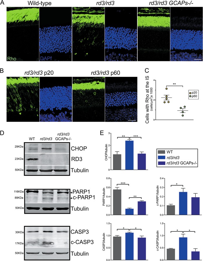
Fig. 6. Rhodopsin mislocalization, ER stress, and apoptosis are early signs of retinal degeneration in rd3 mice that are palliated by GCAPs ablation.
a Retinal sections from wt, rd3/rd3, and rd3/rd3 GCAPs−/− mice at p20 stained with anti-rhodopsin antibody (green) and DAPI (blue). Rhodopsin mislocalization at the perinuclear region and proximal compartments is observed in a number of photoreceptor cells at any given frame of the outer retina in rd3/rd3 mice at p20, but not in wt or rd3/rd3 GCAPs−/− mice this age. Scale bar 20 μm. b, c Representative images and quantification of cells that present rhodopsin mislocalization at p20 versus p60 in rd3 mice, expressed per unit area (n = 5 rd3/rd3 mice at p20; n = 4 rd3/rd3 mice at p60]. Unpaired t-test p20 versus p60, P = 0.0058**). d Levels of CHOP; full length and p17 kDa fragment of cleaved caspase 3; and full length and 89 kDa fragment of cleaved PARP1 proteins in retinal extracts from wt, rd3/rd3, and rd3/rd3 GCAPs−/− at p20. Note that the full-length casp3 and c-casp3 signals were obtained from different exposure conditions of the same membrane, given that c-casp3 represents a small percentage of full-length casp3. No RD3 protein was observed in rd3/rd3 or rd3/rd3 GCAPs−/− extracts. e Six independent experiments were performed to determine CHOP expression levels (wt versus rd3/rd3, P = 0.0012**; and rd3/rd3 versus rd3/rd3 GCAPs−/−, P = 0.0001***). Three independent experiments were performed to determine PARP1 and c-PARP1 levels (PARP1: wt versus rd3, P = 0.001***; and rd3/rd3 versus rd3/rd3 GCAPs−/−, P = 0.0018**); (c-PARP1: wt versus rd3, P = 0.026*; and rd3/rd3 versus rd3/rd3 GCAPs−/−, P = 0.37 NS). Three independent experiments were performed to determine casp3 and c-casp3 levels (casp3: wt versus rd3, P = 0.035*; and rd3/rd3 versus rd3/rd3 GCAPs−/−, P = 0.05*); (c-casp3: wt versus rd3, P = 0.026*; and rd3/rd3 versus rd3/rd3 GCAPs−/−, P = 0.031*).

