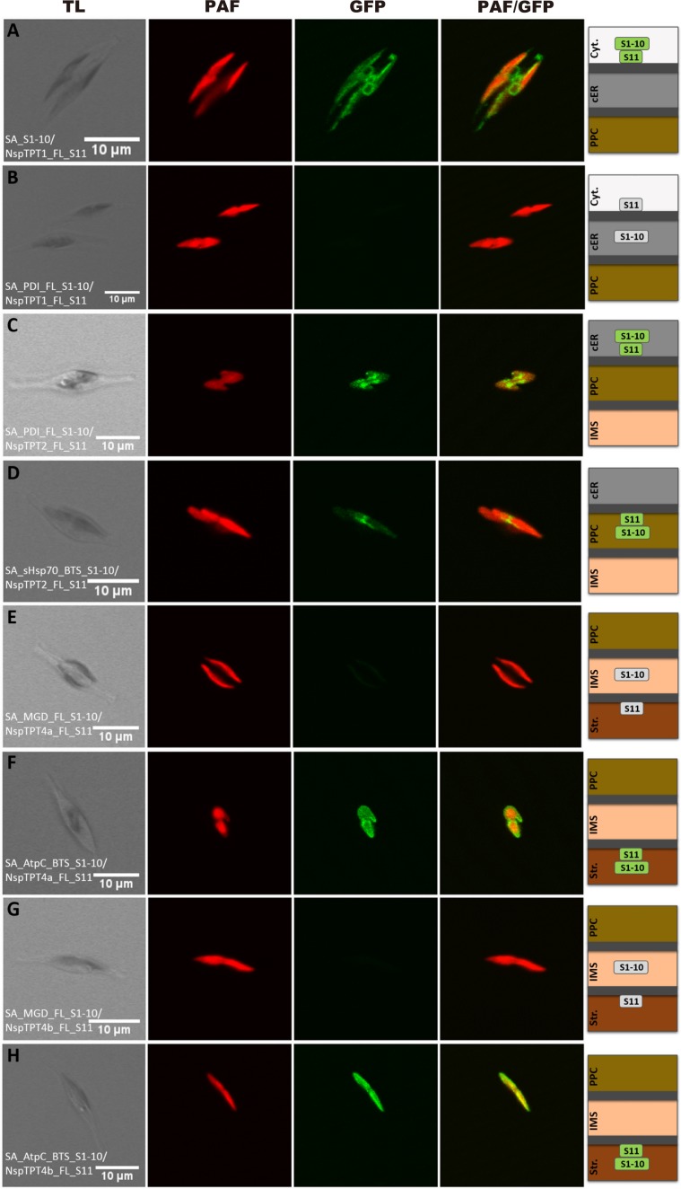
Figure 1.
Self-assembling GFP analysis of non-photosynthetic Nitzschia sp. plastid TPT homologues in Phaeodactylum tricornutum. Simultaneous expression of NspTPT1 fused to GFP(S11) with a cytosolic GFP(S1–10) led to a cER-characteristic fluorescence pattern (A). No fluorescence could be observed after expression of NspTPT1:GFP(S11) with the ER/cER marker (PDI) fused to GFP(S1–10) (B). Fluorescence signals were obtained upon expression of the NspTPT2:GFP(S11) fusion proteins with both the ER/cER- (C) and the PPC-marker fused to GFP(S1–10) (D). Both NspTPT4a:GFP(S11) (E) and NspTPT4b:GFP(S11) (G) did not show a fluorescence signal when expressed with the IMS marker MGD1 fused to GFP(S1–10), whereas a clear GFP signal circling the plastid autofluorescence could be observed when NspTPT4a:GFP(S11) (F) and NspTPT4b:GFP(S11) (H) were expressed with the stromal-targeted AtpC:GFP(S1–10). TL, transmitted light; PAF, plastid autofluorescence; GFP, enhanced green fluorescent protein; PAF/GFP, overlay of plastid and GFP fluorescence; Cyt., cytosol; cER, chloroplast endoplasmic reticulum; PPC, periplastidal compartment; IMS, intermembrane space; Str., plastid stroma; scale bar represents 10 µm.

