
Figure 1.
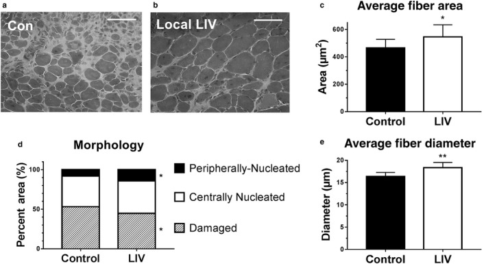
Locally applied low‐intensity vibration (LIV) enhances myofiber growth following laceration muscle injury in mice. Gastrocnemius muscles were lacerated and subjected to local LIV at 90 Hz and 0.2 g in daily bouts lasting 30 min for 14 days. Muscle fiber morphology was assessed in cryosections from the injured portion of the muscle. (a, b) Representative images of hematoxylin and eosin‐stained sections (scale bar = 50 m, 40× magnification). (c) Cross‐sectional area of myofibers, (d) percent area of injury that consists of peripherally nucleated fibers, centrally nucleated fibers, or damaged tissue, and (e) minimum diameter of myofibers was quantified in five 40x fields per muscle in hematoxylin and eosin‐stained sections. N = 3 replicates per group in two separate experiments for a total of N = 6 per group. Data are presented as mean ± SD. *p < .05, **p < .001