Figure 2.
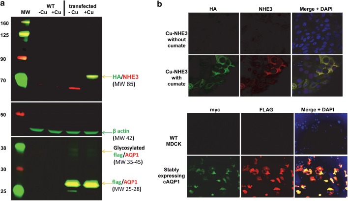
Confirmed overexpression of NHE3 and AQP1. (a) western blot analysis confirming cumate-inducible NHE3 expression (detected by anti-HA and anti-NHE3 antibodies) and AQP1 expression (via anti-flag and anti-AQP1 antibodies). β-actin is provided as a loading control. Molecular weight (MW) markers are labeled to the left of the blots. (b) Immunofluorescent microscopy of transfected cells. Immunofluorescent microscopy of stably transfected MDCK cells expressing cumate-inducible NHE3 expression (detected by anti-HA and anti-NHE3 antibodies) and AQP1 expression (via anti-flag and anti-myc antibodies). Dapi indicates nuclear staining.

