Figure 1.
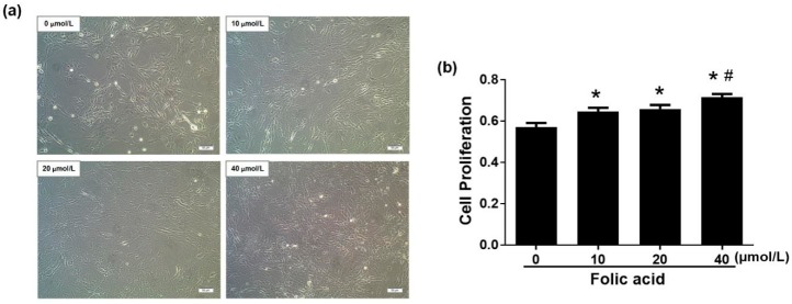
Folic acid increased cell proliferation in primary astrocytes. Primary cultures of rat astrocytes were incubated for 12 days with various concentrations of folic acid (0–40 μmol/L). (a) Cell morphology observed by light microscopy. Scale bar = 100 μm. (b) Bar graph of cell proliferation rates determined by the CellTiter 96® AQueous One Solution Cell Proliferation Assay. The plotted values represent the mean ± SEM values of three experiments. * p < 0.05 compared with the folic acid-deficient group (0 μmol/L), # p < 0.05 compared with the normal-folic acid group (10 μmol/L).

