Figure 6.
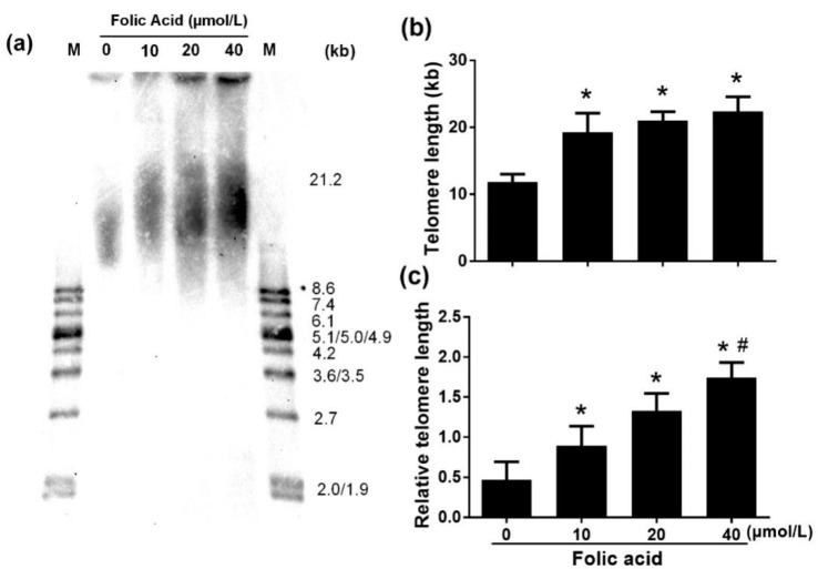
Folic acid inhibited telomere attrition. Primary astrocytes were incubated as described in Figure 1. (a) Mean telomere restriction fragments (TRF) detected by southern blot analysis. (b) Bar graph of southern blot densitometric analysis of mean TRF for genomic DNA. (c) Bar graph of relative telomere length determined by qPCR. The plotted values represent the mean ± SEM values of three separate experiments. * p < 0.05 compared with the folic acid-deficient group (0 μmol/L), # p < 0.05 compared with the normal-folic acid group (10 μmol/L).

