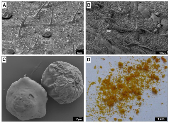
Figure 1.

Scanning electron micrograph showing the distribution of different type of trichomes (FT: fibrous trichomes; PGT: peltate glandular trichomes; CGT: capitate glandular trichomes) on the surface of leaf (A), bracteole blade (B). The scanning electron (C) and light microscopy (D) image of ripe lupulin glands.
