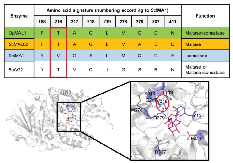
Figure 1.
Amino acid signature of yeast α-glucosidases, including B. adeninivorans AG2 (upper panel) and their designation on the three-dimensional (3D) structure of S. cerevisiae isomaltase IMA1 in complex with isomaltose (RCSB Protein Data Bank, PDB: 3AXH [29]) (lower panel). Location of Val216 in the structure is marked with a red circle.

