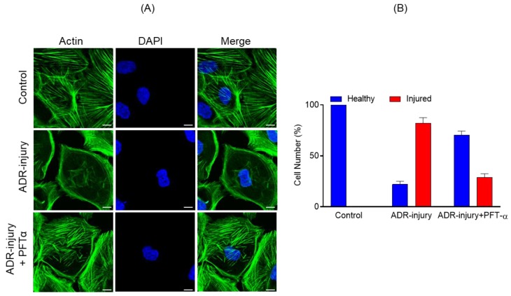
Figure 5.
(A) Podocytes treated with ADR (0.25 μg/mL) for 48 h showed significant actin cytoskeleton damage (green) with accumulation of actin stress fibers at the cell periphery. In contrast, significant recovery of actin cytoskeletal organization was noted in Pifithrin-α (PFT-α)-treated podocytes. Scale bar = 20 µm. (B) The quantitative analysis showed >50% increase in the number of healthy podocytes with a concomitant decrease in ADR injured podocytes upon PFT-α treatment; in contrast, minimal recovery was observed in the vehicle-treated podocytes.

