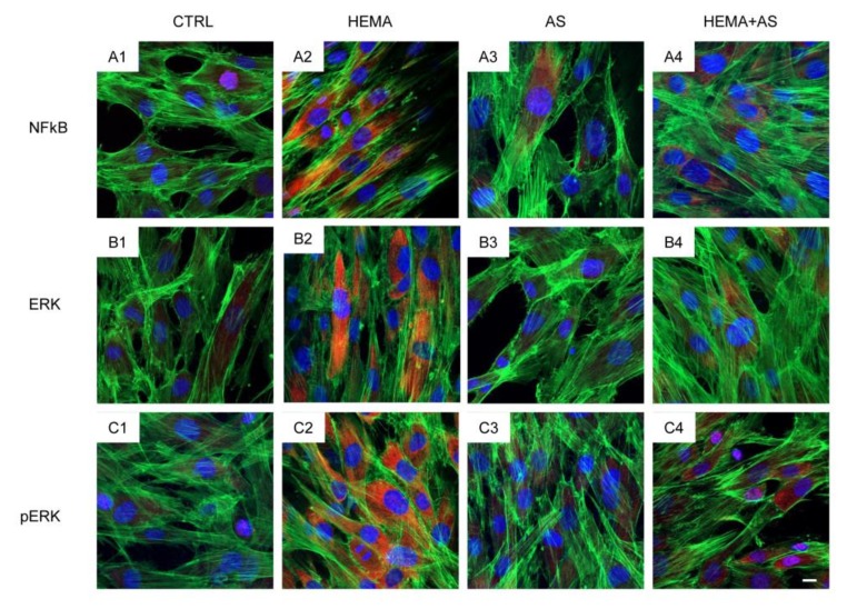
Figure 4.
Untreated cells (CTRL) showed a slight fluorescence signal from NFkB- (A1), ERK- (B1) and pERK-immunostaining (C1). Human DPSCs treated with 2 mM HEMA (HEMA) showed an increased fluorescence levels derived from NFkB-(A2), ERK- (B2) and pERK-immunostaining (C2). Cells treated with ascorbic acid (AS) or HEMA + AS showed fluorescence levels similar to CTRL. Green fluorescence derived from Alexa-phalloidin 488 staining for cytoskeleton actin; red fluorescence derived from Alexa Fluor 568-IGg conjugated secondary antibody to reveal primary antibody against NFkB (A), ERK (B) and pERK (C); blue fluorescence derived from TO-PRO staining of nuclei. Scale bar = 10 µm.

